Microtubule-Modulating Triazolopyrimidines: A Versatile Platform for Therapeutic Discovery
Our laboratory focuses on the rational design and optimization of small-molecule modulators of microtubule dynamics, centered on the 1,2,4-triazolo[1,5-a]pyrimidine (TPD) scaffold. These compounds engage tubulin at distinct binding sites, allowing us to tune cellular phenotypes—from microtubule stabilization to other modulatory effects—through targeted structural modifications.
We apply this versatile chemical platform to develop brain-penetrant therapeutics for neurodegenerative tauopathies, including Alzheimer’s disease, as well as for parasitic infections such as human African trypanosomiasis, with emerging applications in oncology.
Key Approaches
- →Rational design of TPD analogs with tunable cellular phenotypes
- →Optimization of brain penetration and pharmacokinetic properties
- →Structure–activity relationship (SAR) studies around tubulin binding
- →In vivo evaluation in models of tauopathy and parasitic infection
Why Microtubule Stabilization for Tauopathies?
Neurodegenerative tauopathies, including Alzheimer’s disease (AD) and frontotemporal lobar degeneration, are characterized by the accumulation of hyperphosphorylated tau protein that detaches from microtubules. This loss of tau’s normal microtubule-stabilizing function disrupts axonal transport, promotes axonal dystrophy, and contributes to synaptic dysfunction and neuronal death.
While recent Aβ immunotherapies have advanced the field, there remains a critical need for therapies that directly address tau pathology and microtubule dysfunction. The concept of microtubule stabilization as a therapeutic strategy for tauopathies was pioneered approximately 15 years ago by Professors Carlo Ballatore, Kurt Brunden, Virginia Lee, and the late John Trojanowski. Our laboratory continues to build on this foundation, developing brain-penetrant microtubule-stabilizing agents to compensate for the loss of endogenous tau function and restore neuronal integrity.
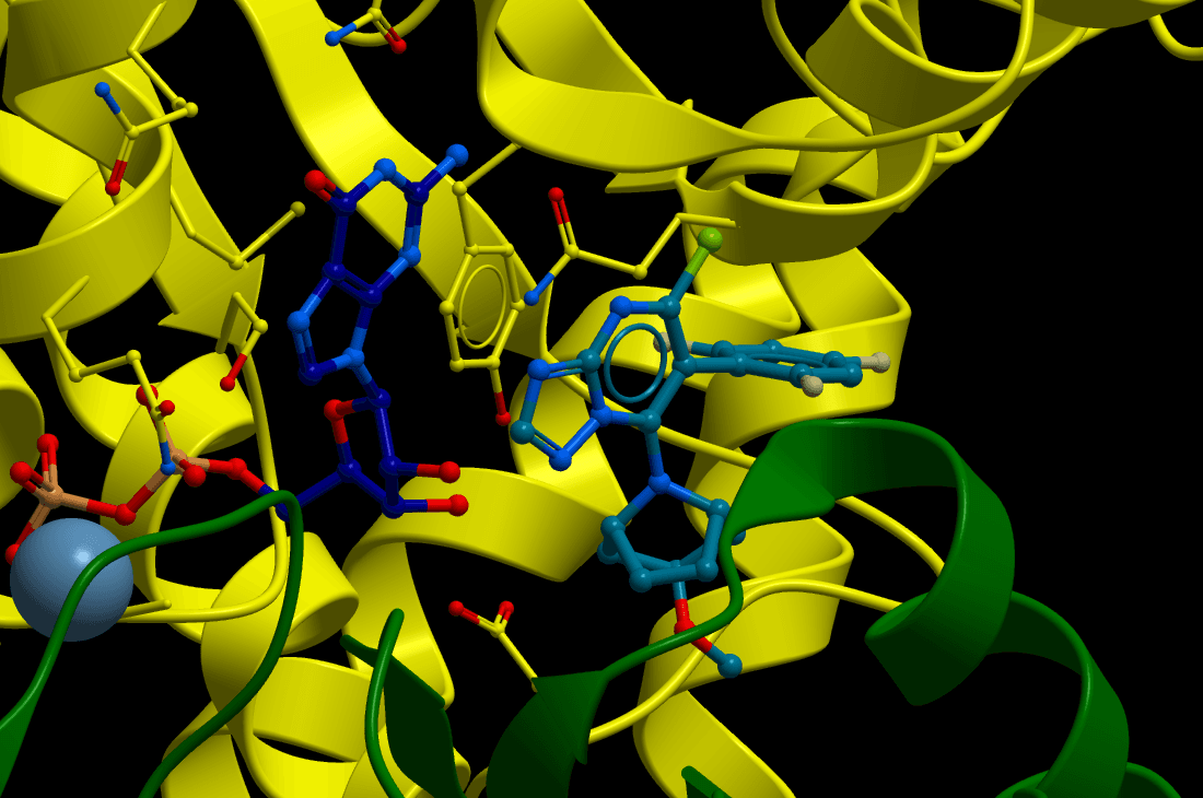
Figure 1. Different mode of action between taxane site binding MT-stabilizers (Epothilone D, TPI-287) and TPDs
Yohannan, Brunden & Alle, Current Topics in Medicinal Chemistry, 2026
Development of Triazolopyrimidine (TPD) MT-Stabilizers
We have focused on the 1,2,4-triazolo[1,5-a]pyrimidine (TPD) scaffold as a synthetically accessible, brain-penetrant platform for microtubule stabilization. These compounds interact with the vinca site on β-tubulin yet promote microtubule stabilization in cells — a mechanism distinct from classical taxane-site binders.
Extensive structure–activity relationship (SAR) studies systematically explored modifications at the C6 and C7 positions of the TPD core. These efforts, reported in Oukoloff et al., J. Med. Chem. 2021and further refined through matched molecular pair analyses and computational studies (Alle et al., J. Med. Chem. 2022), enabled the identification of TPDs with favorable drug-like properties, potent microtubule-stabilizing activity, good brain exposure, and oral bioavailability.
Figure 2. Comparison of selected compounds based on experimental log D7.4 values (triangles) and MT-stabilizing activity (squares) expressed as the average activity in the AcTub assay at 1 and 10 μM normalized to positive control (i.e., 100 nM 5). log D7.4 values were determined via the shake flask method (experiments run by Analiza, Inc.).
CNDR-51997: An Optimized Brain-Penetrant Candidate
Building on the TPD platform, we identified and extensively characterized CNDR-51997, an optimized microtubule-stabilizing candidate with excellent brain penetration and a favorable safety profile.
Figure 3. CNDR-51997 increased the stable microtubule (MT) marker acetylated-tubulin (AcTub) in QBI293 cells and in okadaic acid (OA)-treated primary mouse cortical neurons with MT deficits. (A) Structure of CNDR-51997. (B) CNDR-51997 at 0.3 and 1 μM increased AcTub levels relative to vehicle treatment in QBI293 cells, whereas the compound did not alter total α-tubulin. (C) A volcano plot representing tandem mass tag (TMT)-based quantitative proteomics analysis of QBI293 cells treated with vehicle compared to 10 μM CNDR-51997 shows that compound treatment resulted in a significant >2-fold increase in expression of only the axonemal dynein light chain domain-containing protein 1 (gene name: AXDND1), as indicated by the arrow. Green shading indicates increased and rose shading decreased expression after CNDR-51997 treatment. (D) Primary mouse cortical neurons treated with OA in vehicle only (+OA + Veh) show reduced MT (α-tubulin) staining compared to cultures without OA (−OA). The combined addition of OA and CNDR-51997 at either 1 μM (+OA + 1 μM 51997) or 10 μM (+OA + 10 μM 51997) increased MT staining, more closely resembling neurons cultured in the absence of OA. (E) Quantification of triplicate analyses of primary neuron cultures in the absence or presence of OA, with or without CNDR-51997 addition, as depicted in (D). Error bars represent standard error of the mean (SEM), with p values from one-way ANOVA with Tukey’s multiple comparisons test.
Preclinical Efficacy in Alzheimer’s Disease Models
In detailed studies published in Yao et al., Alzheimer’s & Dementia 2024, CNDR-51997 demonstrated robust efficacy in two complementary transgenic mouse models:
Twice-weekly low-dose administration significantly reduced soluble and insoluble Aβ42, decreased plaque burden, and lowered APP and BACE1 levels — suggesting interruption of a feed-forward cycle between plaques and microtubule disruption.
Three-month intermittent dosing reduced tau pathology, attenuated axonal dystrophy (electron microscopy), decreased neuroinflammation (microgliosis & astrogliosis), and showed a trend toward preserved hippocampal neurons.
Figure 4. A twice-weekly dose of CNDR-51997 as low as 1 mg/kg significantly reduced insoluble and soluble Aβ42 in 5XFAD mice. Male and female 5XFAD mice (1.5 months old) were treated with vehicle or CNDR-51997 at 1, 2, 3, or 5 mg/kg twice-weekly for 4 weeks, followed by ELISA analysis of cortical and hippocampal insoluble and soluble Aβ42 levels. (A) Insoluble cortical Aβ42, (B) insoluble hippocampal Aβ42, (C) soluble cortical Aβ42, and (D) soluble hippocampal Aβ42. Error bars represent standard error of the mean (SEM) with p values determined by comparison of vehicle and CNDR-51997 at each dose using a two-tailed t test.
Figure 5. Treatment of PS19 mice with CNDR-51997 significantly reduced tau pathology. The amount of insoluble total tau (A), AT8-positive phospho-tau (B), and acetylated K280 (AcK280) tau (C) in the brains of PS19 mice after treatment with vehicle or 3 mg/kg CNDR-51997 twice-weekly for 3 months was quantified by immunoblot analyses (also see, Figure S6). The amount of insoluble (D) and soluble (E) brain tau in the PS19 mice was also determined by ELISA. Finally, the amount of tau pathology was assessed by immunohistochemical staining of PS19 mouse brain sections with the MC1 antibody that binds misfolded tau (F), with six bregma levels analyzed for each study mouse and the mean values plotted for each mouse relative to the mean of vehicle-treated mice. The mean percentage area occupied by MC1-positive pathology in PS19 mice varied with bregma level, with mean values in the vehicle group ranging from 47% to 71%. Error bars represent standard error of the mean (SEM) with p values determined by comparison of vehicle and CNDR-51997 treatment using a two-tailed t test.
Our lead candidate is currently advancing through NIH-funded IND-enabling studies, scheduled for completion in July 2027. These studies include multi-kilogram scale-up synthesis, stability and formulation development, as well as comprehensive toxicology and safety pharmacology evaluations.
Expanding the Platform: Repurposing for Parasitic Diseases
Because microtubules are essential in all eukaryotes, we have explored whether our TPDs library developed for central nervous system indications could be repurposed against neglected tropical diseases (work in collaboration with the Caffrey lab, Skaggs School of Pharmacy, UCSD).
Human African Trypanosomiasis (HAT)
Screening of 123 TPD congeners enabled a robust QSAR model. Prioritized compounds rapidly reduced parasitemia in Trypanosoma brucei-infected mice. Once-weekly dosing significantly extended survival, highlighting potential for stage 2 HAT (CNS involvement).
Figure 6. Kaplan–Meier curves for survival of T. brucei-infected mice treated with TPDs. Female BALB/C mice were infected i. p. with 1×10⁵ T. b. brucei Lister 427 parasites. On day 2 post-infection, when parasitemia was established, mice were divided into groups of five and treated with an i. p. injection of (A) 4 mg/kg pentamidine (green), or 5 mg/kg (orange), 7.5 mg/kg (red) or 10 mg/kg (blue) of 3 (B) or 4 (C). Black lines in each panel indicate the survival of infected mice treated with vehicle alone. An additional dose of TPD 3 or 4, as indicated in the text, was administered on day 9 post-infection (▾) when parasitemia was detected. Asterisks (*) indicate the presence of blood parasitemia at the end of the study (days 15 and 16 post-infection for 3 and 4, respectively)
Schistosomiasis
Phenotypic screening within our MT-active library identified potent activity against Schistosoma mansoni. Optimized congeners produce rapid, long-lasting paralysis of adult worms and larvae with substantially improved selectivity over mammalian cells.
Figure 7. (A) Concentration-dependent paralysis of adult S. mansoni after a 5 h exposure to phenylpyrimidine compounds 25, 28, and 29 at concentrations ranging from 0.31 to 10 μM. (B) S. mansoni adult worm motility remains suppressed even after removal of 25, 28, or 29. Parasites were preincubated with 0.5% DMSO or 2 μM of test compound for 5 h. Just prior to exchanging the incubation volume six times (Time 0), worm motility was measured by WormAssay and then again at 24 and 48 h after the exchange
KEY PUBLICATIONS
Monti et al. — Congeners Derived from Microtubule-Active Phenylpyrimidines... Schistosoma mansoni. ACS Infect. Dis., 2021